Imaging Laboratory

Laser-scanning confocal microscopes
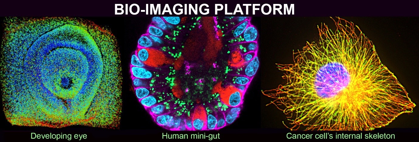
With additional funding support from UKRI, our microscopes include the very latest technology in confocal microscopy, the facility contains two laser-scanning confocal microscopes the Zeiss LSM 980 Airyscan2 with super-resolution capability and the Zeiss Observer 7, an advanced widefield fluorescence microscope for sensitive, fast, live imaging in four dimensions. A Zeiss LSM510 META confocal and TriM Scope II multi-photon microscope is also available.
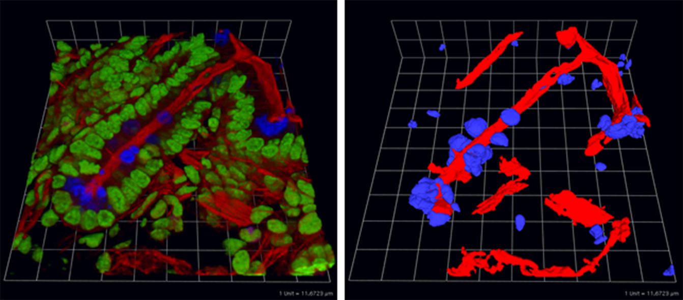
The newly installed HIVE data storage system from Acquifer together with the Aivia image analysis software with Artificial Intelligence capability (DRVISION Technologies) and Huygens deconvolution software (Scientific Volume Imaging) will provide critical support for the microscopes and the latest technology in 4D-image analysis. Together, the new cutting-edge microscopes and image analysis systems will enable multiple researchers at UEA and across the NRP to gain unprecedented data from super-resolution microscopy and sensitive, fast, live imaging across scales, from single molecules and cellular structures to 3D organoids, tissues and whole organisms.
TriM Scope II multi-photon microscope
This microscope is used for either deep-tissue imaging or Fluorescence Lifetime IMaging (FLIM). It incorporates both an upright and an inverted stand that share the same scan-head in order to accommodate virtually any sample. The 8 detectors include a 16-channel PMT array for very fast, lifetime imaging.

Wide-field, CCD camera set-ups
There are four microscopes for acquisition of widefield, fluorescence images. Three microscopes (one upright and two inverted) are equipped with monochrome cameras and motorized stages together with the software to obtain multi-channel, time-series and Z-stack data; the fourth microscope is a stereomicroscope with a colour CCD camera.

Note: There are also two POC (Perfusion, Open and Closed cultivation) chambers and two Ludin chambers that can be used on all the inverted microscopes to allow perfusion as well as temperature and CO2 regulation.
Analysis Suite

The analysis suite on Floor 01 of building 5 contains three workstations each having 18” LCDs, dual 2.4 GHz processors, 64 GB RAM, 2x 1 TB of hard disk space (configured in RAID 0) with either GeForce GTX-780 (3 GB VRAM) or Quadro P4000 (8 GB VRAM) graphics cards. These computers are equipped with the software to carry out time-series analysis, object tracking through time or space, 3D reconstruction and image deconvolution (Andor iQ , AxioVision, Fiji/ImageJ, FLIMfit, Huygens, ilastik, ImSpectorPro, ImagePro, Volocity and Zen Blue). With these software packages comes the ability to export files in numerous formats.
The PCs are also loaded with standard Microsoft Office software and Adobe Photoshop to allow image refinement and incorporation into documents and presentations.
Printing facilities include 2 photoquality colour inkjet printers, 1 colour laser printer and 1 monochrome laser printer.
)
)